Home

Begradigen Cordelia Tasche yeast cells in stool Überraschung Morbidität wie oft

red and white blood cells, and yeast in urine sediment | Medical  Laboratories
red and white blood cells, and yeast in urine sediment | Medical Laboratories

Budding Yeast Cells With Pseudohyphae Stock Photo - Download Image Now -  Thrush - Yeast Infection, 2015, AIDS - iStock
Budding Yeast Cells With Pseudohyphae Stock Photo - Download Image Now - Thrush - Yeast Infection, 2015, AIDS - iStock

Direct microscope stool examination, red arrow show Candida budding,... |  Download Scientific Diagram
Direct microscope stool examination, red arrow show Candida budding,... | Download Scientific Diagram

PDF] DARK-FIELD MICROSCOPE STOOL ANALYSIS – ITS ROLE IN DIAGNOSIS OF YEAST  OVERGROWTH IN GUT | Semantic Scholar
PDF] DARK-FIELD MICROSCOPE STOOL ANALYSIS – ITS ROLE IN DIAGNOSIS OF YEAST OVERGROWTH IN GUT | Semantic Scholar
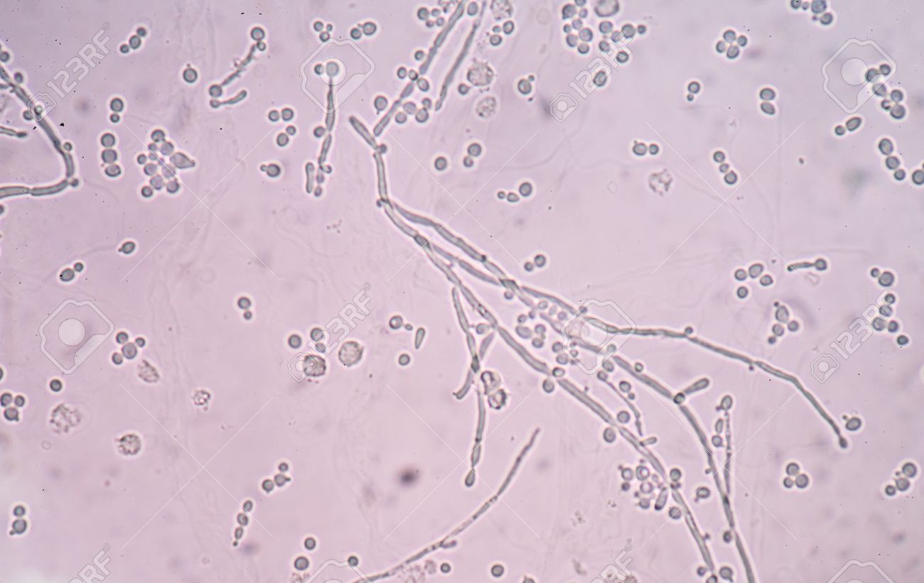
Branching Budding Yeast Cells With Pseudohyphae In Urine Sample Fine With  Microscope. Stock Photo, Picture And Royalty Free Image. Image 44502230.
Branching Budding Yeast Cells With Pseudohyphae In Urine Sample Fine With Microscope. Stock Photo, Picture And Royalty Free Image. Image 44502230.

Entamoeba Coli Protozoa In Stool Exam Stock Photo - Download Image Now -  Dinoflagellate, Bacterium, Microscope - iStock
Entamoeba Coli Protozoa In Stool Exam Stock Photo - Download Image Now - Dinoflagellate, Bacterium, Microscope - iStock
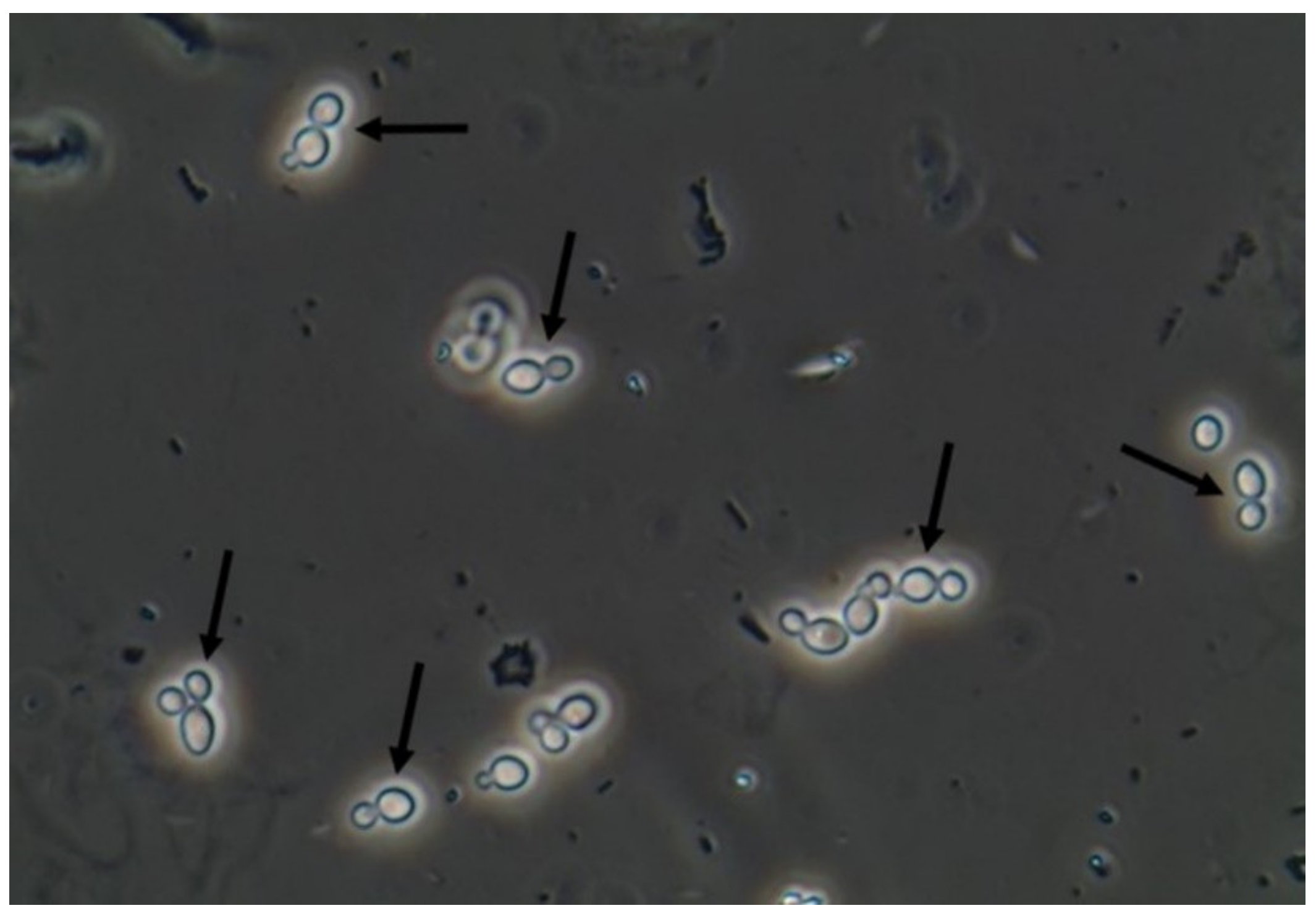
JoF | Free Full-Text | Urine Sediment Findings and the Immune Response to  Pathologies in Fungal Urinary Tract Infections Caused by Candida spp.
JoF | Free Full-Text | Urine Sediment Findings and the Immune Response to Pathologies in Fungal Urinary Tract Infections Caused by Candida spp.

Bacteria and Fungus Microscopy at Various magnifications 400X, 800X and  1600X - YouTube
Bacteria and Fungus Microscopy at Various magnifications 400X, 800X and 1600X - YouTube

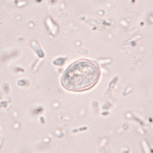
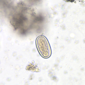
CDC - DPDx - Artifacts

CDC - DPDx - Artifacts

CDC - DPDx - Artifacts

Yeast cells in urine microscopy. - YouTube

Fungal element in stool | Fungal infection | fungal under microscope | how  to identify fungal - YouTube
Fungal element in stool | Fungal infection | fungal under microscope | how to identify fungal - YouTube

Dimorphic fungus - Wikipedia

Wet Mount Proficiency 2006A Critique

117 Cells Fungi Microscopy Stock Photos - Free & Royalty-Free Stock Photos  from Dreamstime
117 Cells Fungi Microscopy Stock Photos - Free & Royalty-Free Stock Photos from Dreamstime

Microscopic Morphologic Features of Yeast | Medical Laboratories

Dimorphic Fungal Coinfection as a Cause of Chronic Diarrhea and Pancolitis

numerous yeast cell seen in patient's stool 🌿 | Yeast, Celestial bodies,  Cell
numerous yeast cell seen in patient's stool 🌿 | Yeast, Celestial bodies, Cell

Wet Mount Proficiency 2006A Critique

Fungal Diseases
Fungal Diseases

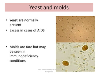
Routine examination of stool

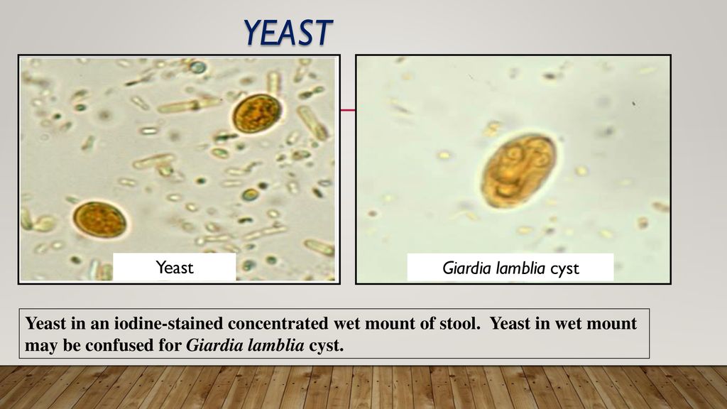
Artifacts Medical parasitology lab. Faculty of health sciences - ppt  download
Artifacts Medical parasitology lab. Faculty of health sciences - ppt download